
Here you find showcase research results of the Virtual Institute.
Overviev:
X-ray imaging of liquids in porous materials enhanced
Partners involved: EMPA, TUM, KIT-IMT
Standard, attenuation-based X-ray imaging of liquids in porous materials requires adding to the liquid a “contrast agent”, i.e., a substance containing e.g. Cs, Ba or I, to increase its linear attenuation coefficient. This requirement is a strong limitation for imaging chemically reactive liquid transport processes, e.g., water transport in concrete and in polymer electrolyte membrane fuel cells (PEMFC) during operation. A new approach to X-ray imaging of weakly attenuating liquids, e.g. pure water, in materials with pore space dominated by small pores (below hundreds/tens of microns) is based upon laboratory-scale Talbot-Lau interferometry. Using a polychromatic tube source, imbibition of concrete samples with pure water was imaged in real time. This approach does not require any contrast agent but exploits the intrinsic, sub-pixel scale X-ray multi-refraction due to the micro structural heterogeneity of the materials themselves. The approach has the potential to become complementary to neutron and magnetic resonance imaging of water in porous materials.


X-ray radiographs at start and after one hour of the capillary uptake. (a) and (b) dark-field radiographs. Brighter pixel values correspond to larger scattering strength. (c) and (d) attenuation radiographs. Brighter pixel values correspond to larger X-ray attenuation. The bright horizontal lines visible towards the bottom in (c) and (d) are artifacts created by evaporation from the water surface during the experiment.

Wetting front positions (red lines) overlaid on top of a central region of interest of the last absorption radiograph, for the three different samples. For each sample, the first wetting front is plotted at approximately 2.9 min after water immersion. The time gap between two successive fronts is about 4.6 min.
Enhancing X-ray imaging of liquids in porous materials, R. Kaufmann, F. Yang, F. Prade, M. Griffa, I. Jerjen, C. di Bella, J. Herzen, A. Sarapata, F. Pfeiffer, P. Lura, A. Neels; Digital Industrial Radiology and Computed Tomography (DIR 2015), 22.-25.6.2015, Belgium, Ghent, www.dir2015.ugent.be
F. Yang, F. Prade, M. Griffa, I. Jerjen, C. Di Bella, J. Herzen, A. Sarapata, F. Pfeiffer, P. Lura, Dark-field X-ray imaging of unsaturated water transport in porous materials, Appl. Phys. Lett. 105, 154105 (2014). DOI: 10.1063/1.4898783
Enlarging computed tomography realized with Taille-lenses
Partners involved: KIT-IMT, KIT-IPS, HZG, TUHH, SFB 896, ESRF-BM01
In a conventional medical tomography setup the X-ray source and the detector rotate around the patient. However in the technical field source and detector mostly do not move as the sample is rotated around its axis. When using an X-ray lens to image the sample to the detector the method is called nano-CT. Such a setup has been established at PETRA III-P05 using Taille-lenses designed at KIT-IMT. The example shows a photonic glass consisting of zirconium oxide spheres. The measurement has been carried out in cooperation with the TUHH in the scope of the SFB 986 (see about). A resolution of 200 nm per line and space was achieved in this measurement.

3D-view of a section from a CT-measurement of zirconium oxide spheres (ca. 15 µm edge length) ©02, ©04
Rolled X-ray mirror optics assessed
Partners involved: KIT-IMT, KIT-IPS
Rolled X-ray Mirror Optics (RXMOs) are optics similar to the Rolled X-Ray Prism Lenses (RXPLs) in so far as they are also produced by rolling a thin polyimide foil around a winding core. Gold or other meterials can be vapour-deposited on the mirroring surface of the foil to increase reflectivity. There are spacers placed regularly on the foil, resulting in a small air gap between windings after the rolling. X-rays entering these air gaps are reflected and focused in the focal plane. Main challenge in producing these foils is the precision needed during the rolling process to ensure small angle deviations in the placement of the foil relative to the optical axis. First Experiments at the synchrotron source ANKA-TOTO showed the existence of a focal point, although the spectral intensity gain was low due to technical shortcomings.


Rolled X-ray mirror optics: sketch of the inner buildup (left) and radiograph showing the foil layers with spacers in between (right)
Full field microscopy using Taille-lenses
Partners involved: KIT-IMT, KIT-IPS, HZG, ESRF-BM01
Setups for X-ray full field microscopy using refractive compound lenses (CRLs) with varying aperture (=Taille-lenses) and rolled X-ray prism lenses (RXPLs) as condensers have been implemented at the synchrotron sources ANKA-TOTO, PETRA III-P05 and ESRF-BM01. The condenser illuminates the sample; the imaging optics creates an enlarged image on a scintillator. The scintillator converts the X-rays to visible light which can be detected using a CCD-detector with suitable optics. A 45°-mirror protects the detector from the direct X-rays passing through the scintillator.

Sketch of a full field microscopy setup: the part left of the scintillator is the X-ray optical part, on the right side of the scintillator the visible light part
The setup has been used with photon energies of 17.4 keV and 30 keV. In a field of view of 80 µm x 80 µm lines thinner than 100 nm could be resolved, corresponding to a resolution of better than 200 nm per line and space.


The lower pictures show full field microcopy images of a test pattern consisting of lines with a thickness of 120 nm (left) and 90 nm (right) taken at 17.4 keV at PETRA III-P05; the upper half shows SEM pictures of the same test patterns ©02

Full field microcopy image of a Siemens star at 17.4 keV at PETRA III-P05 ©01
F. Marschall, A. Last, S. Georgi, D. Lamago, O. Markus, V. Nazmov, M. Simon, H. Vogt, J. Mohr, Innovative aperture-optimised refractive lenses for hard X-ray full field microscopy, zur Veröffentlichung eingereicht beim Journal of Synchrotron Radiation, (2014)
Bent X-ray gratings fabricated successfully
Partners involved: KIT-IMT, PSI
X-rays are radiated from an X-ray tube source with large divergence angle. To avoid shadowing effects in grating based X-ray tomography, the gratings lamellae need to be oriented parallel to the ray's direction. This can be achieved, e.g. by bending the substrate (usually a silicon wafer) together with the grating to a radius equal to the distance of the grating from the source point. We realized bent phase gratings as well as the source gratings with periods of 2.4 µm and more, fabricated on a silicon wafer, 200 µm thick. With such bent gratings the field of view in phase contrast imaging was doubled for standard setups.


X-ray gratings on a bent silicon wafer used in phase contrast imaging at tube sources (left); phase contrast image of a screw: image taken with a non-bent grating (up), large field of view due to the used bent grating (down) ©01, ©03
„Sun rays“ stabilize X-ray grating structures
Partners involved: KIT-IMT
The period of the X-ray gratings which are used in phase contrast imaging is only a few micrometers. For the source grating (G0) and the absorption grating (G2) the lamella height needs to be more than hundred micrometers in case of design energies of more than 40 keV. Otherwise the absorption is not high enough to achieve a good visibility. Those gratings are fragile and it is a challenge to fabricate them with reasonable quality. Due to resist shrinkage and different process temperatures the gratings are exposed to stress. Moreover during convection drying after the wet development, capillary forces lead to agglutination of the lamellas. To overcome this problem and to stabilize the gratings mechanically resist bridges were introduced. Although agglutination could be avoided, the connected mesh cracked randomly in certain distances. Cracking could be reduced drastically by connecting the lamellas by so-called „sun rays“. These are pin like structures with a diameter of 2 to 3 µm which are fabricated by a 45° exposure. They penetrate several lamellas from top to bottom stabilizing the lamellas as well. Compared to the bridges they do not influence the visibility locally. This improved the grating quality remarkably.

X-ray gratings with bridges (left) and „sun rays" (right) ©01
The gratings quality has been improved in addition by freeze drying the developed resist structures. As the solvent will not be evaporated in the drying process but frozen and sublimated, no capillary forces are generated. This avoids agglutination of the grating structures without any stabilizing structures to a certain height (up to an aspect ratio of 20).
J. Mohr, A. Last, V. Nazmov, M. Simon, Th. Grund, J. Kenntner, Resiststruktur zur Herstellung einer röntgenoptischen Gitterstruktur, Patent WO2012055495, Anmeldedatum 13.11.2011, (2011)
Partners involved: KIT-IMT, HZG
The direction of X-rays is nearly not changed when X-rays pass matter. Thus it is difficult to make lenses for X-rays: X-rays experience a bit of absorption when passing through a lens, but they are hardly focused. When Wilhelm Conrad Röntgen discovered X-rays, he thought it was impossible to make refractive X-ray lenses. Refractive X-ray lenses can be relized, when the refracting surfaces have very small radii of curvature and when the X-rays pass hundreds of these lenses aligned in a row. Moreover focusing lenses for X-rays have to have a concave shape instead of a convex shape (as for visible light).
Until now at KIT-IMT imaging Refracting Compound X-ray Lenses (CRLs) have been fabricated as long rows of equal lens elements. The fabrication process is deep X-ray lithography in negative resists like SU-8.
Refracting Compound X-ray Lens; in this SEM-image X-rays pass through the lens from bottom to top, but only the crossed section of the parabolic shaped par tof the lenses is used as lens ©01
Further development at KIT-IMT led to so called "Taille"-lenses. In this type of CRLs the individual lens elements of one CRL have varying apertures along the optical axis. Extensive numerical simulations showed that the optical properties of CRLs can be improved when the lenses have larger local apertures at the lenses ends than near it's centre. As such lenses have kind of waist, we named them "Taille"-lenses after the German word for waist. The optical properties are improved, because the lenses for a given focal length can be a bit shorter than CRLs with constant apertures. As a consequence, they have a larger effective aperture and thus can achieve higher resolution in imaging applications. Also the field of view is widened, allowing for larger samples. Taille-lenses show a more homogeneous intensity distribution over the field of view.

Comparison of the simulated intensity distributions homogeneity when imaging with a conventional CRL (left) and a Taille-CRL (right) ©01
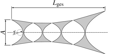
Exaggerated sketch of the shape of a Taille-CRL ©01
With such lenses we imaged 100 nm wide lines (thus a resolution of 200 nm per line and space) in a field of view of 80 µm x 80 µm at PETRA III-P05. Theoretically resolutions of 50 nm per line and space respectively fields of view of up to 500 µm should be possible.
F. Marschall, A. Last, S. Georgi, D. Lamago, O. Markus, V. Nazmov, M. Simon; H. Vogt, J. Mohr, Innovative aperture-optimised refractive lenses for hard X-ray full field microscopy, zur Veröffentlichung eingereicht beim Journal of Synchrotron Radiation, (2014)
A. Last, F. Marschall, Röntgenlinsenanordnung und Herstellungsverfahren, zur Patentanmeldung eingereicht 19.5.2014
Mobile X-ray detectors built up
Partners involved: KIT-IMT
At many X-ray sources there is no suitable X-ray detector available for alignment and characterization of X-ray lenses. To be able to characterize and use improved X-ray lenses quickly and with good accuracy at whatever source, at KIT-IMT two mobile X-ray detectors have been constructed. In these detectors a LSO-scintillator crystal with 8 mm diameter (can be changed to other types) converts incoming X-rays to visible light. The visible light then is imaged via a 45°-mirror, a microscope objective and a tube lens to a CCD-detector.
The first detector is very compact, because the optical path it folded several times. The detector has external dimensions of 100 mm x 180 mm x 280 mm, a fixed field of view of 1.7 mm x 1.3 mm at an optical resolution of 1.5 µm in the plane of the scintillator.
The second detector is based on a commercial digital camera with interchangeable objectiv lenses. The detector is capable of taking 60 frames per second videos, a helpful feature when aligning lenses or to judge the stability of the X-ray beam. A PCO4000-detector is available and can be used as well. Field of view and resolution depend on the objective used and range from 1.8 mm x 1.2 mm with 0.8 µm resolution and 18 mm x 12 mm with 10 µm resolution.
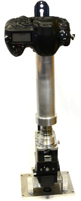
Mobile X-ray detector with interchangeable optics (left) and compact X-ray detector (right) ©01
Increase in photon intensity in diffractometry
Partners involved: KIT-IMT, KIT-IAMWK, Bruker AXS GmbH
Usually the optics in a diffractometer setup consists of a Montel mirror and a beam collimator to achieve a beam spot in the range of several hundred micrometres. The beam collimator is a simple metal tube and only blocks the photons which are not hitting the metal tube's opening. Thus, only a small part of photons are available to illuminate the sample; most of them are absorbed generating unwanted heat. At a diffractometer at Bruker AXS we exchanged the metal tube by a rolled X-ray prism lens (RXPL). With this lens we measured an increase of the integrated photon intensity by a factor of around 18. This allows either to achieve a better signal to noise ratio in the same measurement time or to reduce the measurement time considerably, which is important for commercial X-ray diffractometers.


Diffractogram of a corundum sample (NIST 1976a) taken with an X-ray tube source (Siemens KFL-CU-2K (Cu)). The set-up contained a Montel mirror (Incoatec Montel-p) and a metal tube collimator (left), an RXPL (right) ©01
H. Vogt, A. Last, J. Mohr, F. Marschall, K.-U. Mettendorf, R. Eisenhower and M. Simon, Low-cost Rolled X-ray Prism Lenses to increase photon flux density in diffractometry experiments, Powder Diffraction, available on CJO2014 (2014), DOI:10.1017/S0885715614000177
Rolled X-ray prism lenses have been successfully fabricated
Partners involved: KIT-IMT
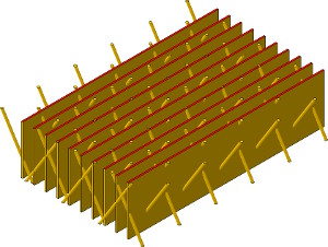
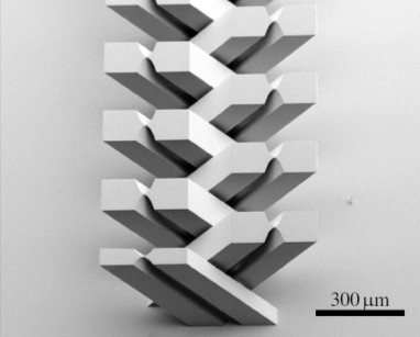
So called Rolled X-ray Prism Lenses (RXPL) open up the possibility to fabricate illumination optics with large aperture. Such lenses have been realized at KIT-IMT for the first time. They are fabricated by winding a thin micro structured polyimide foil, which carries parallel ridges with triangular cross section. Due to the small refractive power of a single ridge, only after passing thousands of ridges the X-rays are refracted to a reasonable focal distance. To keep the loss in X-ray intensity small the penetration length through the ridges should be as small as possible; the edge length of the ridges is in the range of 10 µm only.
To fabricate the lens the foil is cut into a specific shape and subsequently wound around a 125 µm thin glass fibre. The shape of the foil determines the focal distance depending on the photon energy.

Fabrication of the micro structured polyimide foil ©01


Foil and finished RXPL; in the sketch the winding core is omitted ©01
Finished Rolled X-ray Prism Lenses can have diameters between 500 µm to 2 mm, what is quite large for X-ray optics. As shown in the picture above the number of prisms which are transmitted by the X-rays increases from the centre of the lens to the boundary, resulting in an increased refractive angle so all individual beams are pointing to the focal spot. We aim to use these lenses as illumination optics in an X-ray microscope for hard X-rays.
H. Vogt, A. Last, J. Mohr, F. Marschall, M. Kluge, V. Nazmov, K. U. Mettendorf, R. Eisenhower, Refraktive Röntgenlinsen zur Intensitätserhöhung im Spot einer Röntgenröhre, Proc. MikroSystemTechnik Kongress 2013, Aachen, VDE-Verlag, S. 453-456 (2013)